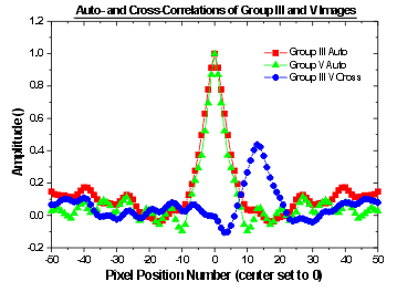
 Fig. 9: Line scans across the dominant peaks of the auto- and cross-corelations of the Group-IV related and Group III related images of Fig. 7 and 6, respectively. The line scans are along the x directions. The red and green curves show the autocorrelations of the Group-III and -V related images, respectively. The central peak with amplitude of 1.0 represents direct overlap of the images, while the smaller peaks are due to the atomic periodicity in the images. The blue curve shows the cross-correlation of the group III and V images. The peak with amplitude 0.4 at near pixel 13 indicates a high degree of correlation between the two images with the image positons are offset by about two atoms in the [001] direction (0 atoms in the [ 1-10] direction). This offset appears to be due to variations in the group-V and -III related images of the defect used to align the images.
[Click on figure to enlarge]