|
1
|
- Imaging methods (microscopy)
- optical bright field
- optical dark field
- electron microscopy
- Non imaging (light scattering)
|
|
2
|
- Gold and silver nanoparticles
- Characteristics
- Strongly absorbing
- Refractive index is a strong function of wavelength
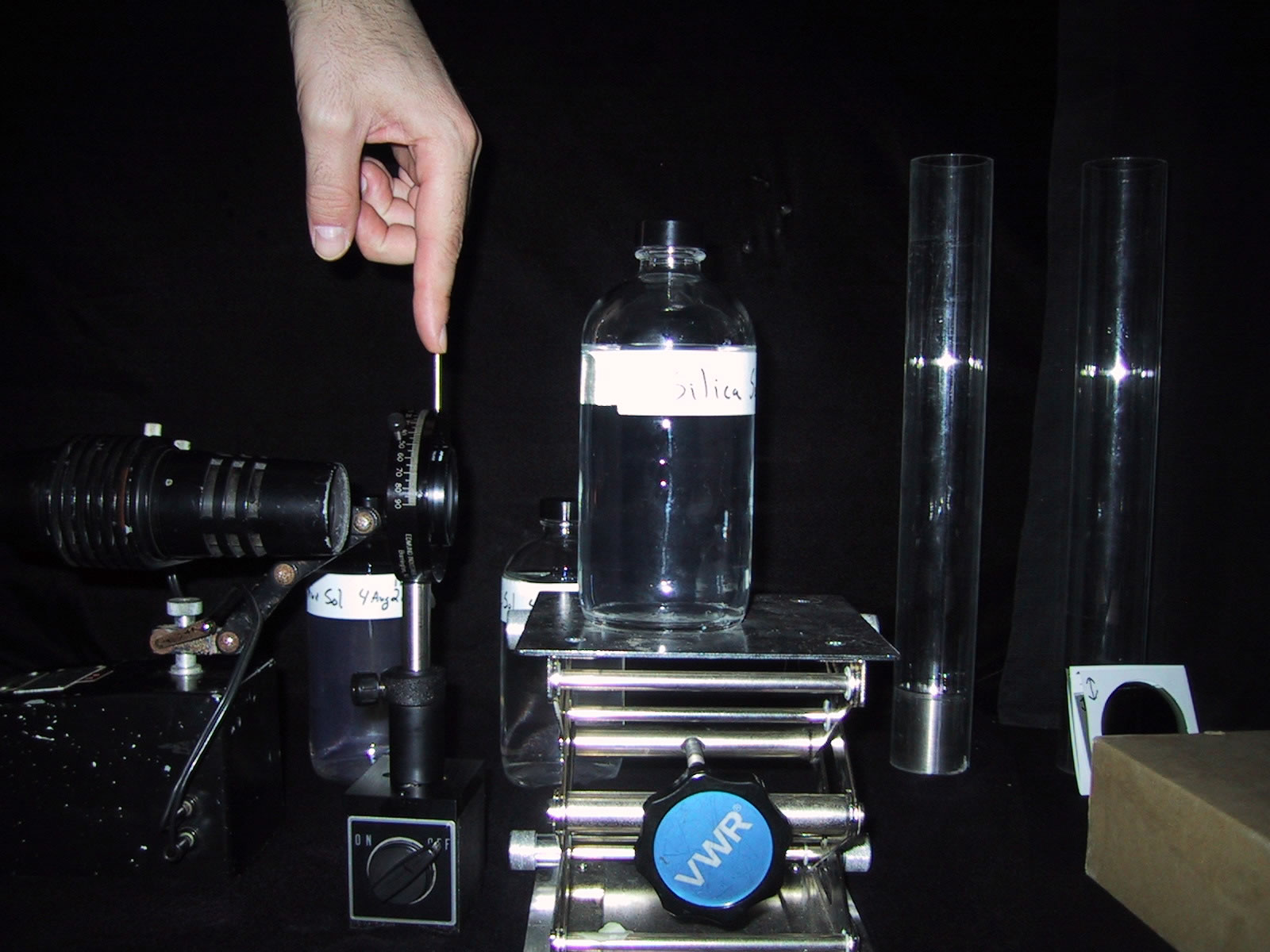
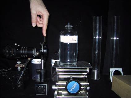
- Experiment
- Preparation
- Visual inspection (color & scattering)
- Absorption spectra
- Polystyrene and sulfur nanoparticles
- Characteristics
- Non-absorbing
- Refractive index is a weak function of wavelength
- Experiment
- Visual inspection (angular scattering, polarization, HOTS)
- Angular scattering
|
|
3
|
- distinguish between color of scattered light and the color of
transmitted light
- single scattering (dilute)
- multiple scattering (concentrated, e.g. milk)
|
|
4
|
- Absorption
- Reflection
- Refraction
- Diffraction
- large objects each effect is distinct
- small objects the difference between the latter three effects is
blurred.
|
|
5
|
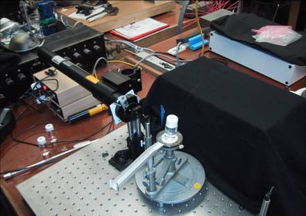
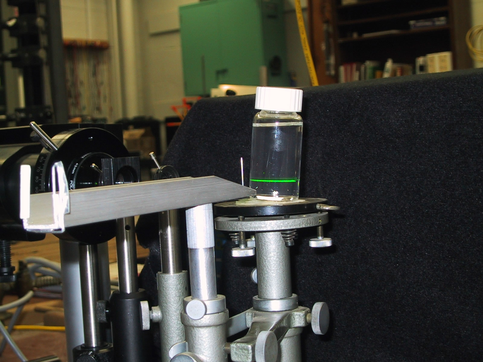
- A collimated light source is the most basic tool for nanoparticle
work. Often called a Tyndall
beam. Named after the 19th
century scientist John Tyndall who studied light scattering in detail.
- The Tyndall effect is the scattered path of light observed in the
suspension. Examples: Milk, smoke, Lake Thunderbird.
|
|
6
|
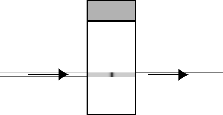
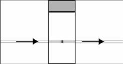
- absorption (light absorbed and converted to heat)
- scattering (light deviated from its original path)
- extinction (light removed from the beam by absorption and scattering)
|
|
7
|
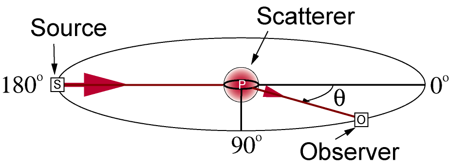
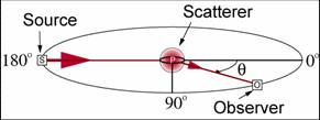
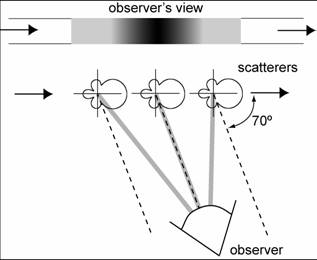
- How is the angle measured?
- Zero is the forward direction, the direction of the undeviated rays
- 180° is backward, rays scattered directly back into the source.
- Note that in the diagram to the
right the scattering angles are
129 ° (180° – 51°) and
138 ° (180° – 42°),
respectively.
|
|
8
|
- the scattering plane is defined by the two rays involved, the
source-particle ray and the particle-observer ray
- The scattering plane is determined by observation, it is not fixed in
space.
- For example, if the observer moves, the scattering plane will move with
the observer
- The scattering plane is useful to define the direction of polarization
of light (parallel and
perpendicular)
|
|
9
|
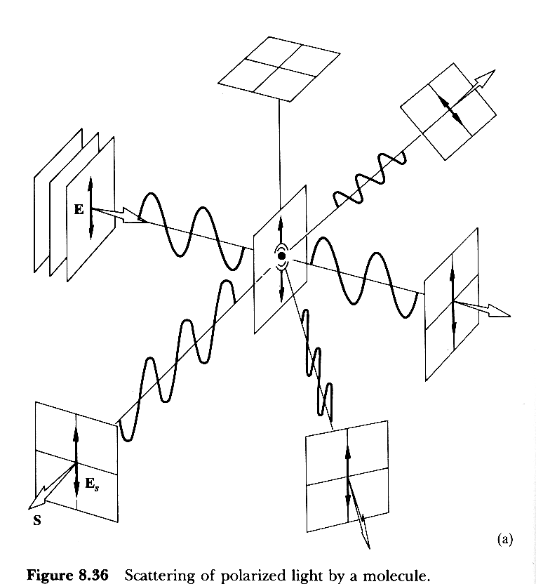
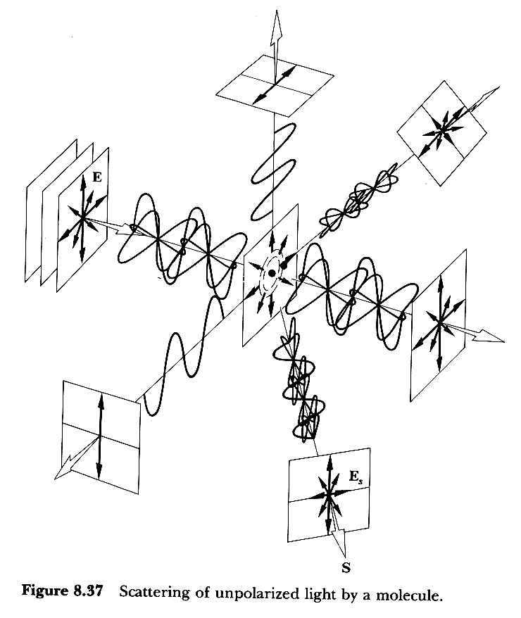
- Lord Rayleigh AKA John Strutt (1871) motivation: Why is the sky blue?
- The particles are treated as electric dipoles
- results:
- I ;1/
λ4 (only true if the refractive index is a weak
function of λ, i.e. not a metal.)
- I ;r6
- scattered light at 90° is linearly polarized perpendicular to the
scattering plane
|
|
10
|
|
|
11
|
- unpolarized source
- Note that 90° scattering
is polarized perpendicular to the scattering plane.
|
|
12
|
- Gustav Mie (1908)
motivation: The colors of
colloidal gold.
- Multipole expansion (EM modes of a sphere)
- electric dipole
- magnetic dipole, electric quadrupole
- magnetic quadrupole, electric octupole
- etc.
- If d < λ/20 then only the first term (dipole) is needed. In this limiting case, Mie’s theory
reduces to Rayleigh’s theory
- small particle limit: Mie à Rayleigh
|
|
13
|
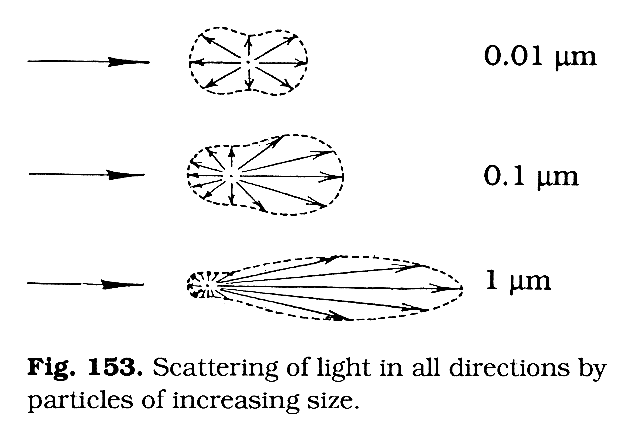
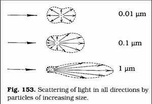
- simple in the Rayleigh limit
- larger sizes show complicated behavior
- general trend is that larger particles scatter more light in the
forward direction.
|
|
14
|
- Efficiency factors: Qsca,
Qabs, Qext
- Plot Qext vs λ for the extinction spectrum
- Qsca and Qabs vs λ show their contribution
to Qext.
- Intensity for perpendicular and parallel polarized light
- Plot I vs θ for the angular intensity dependence for each
polarization.
|
|
15
|
- Craig F. Bohren, Donald R. Huffman, Absorption and Scattering of Light
by Small Particles, (Wiley, 1983). [general reference]
- H. C. van de Hulst, Light Scattering by Small Particles, (Wiley, 1957
& Dover, 1981). [general reference]
- Marcel Minneart, The Nature of Light & Color in the Open Air,
(Dover, 1954) . [great pictures
and explanation of atmospheric phenomena]
- Marcel Minneart, Light & Color in the Outdoors, (Springer,
1992). [great pictures and
explanation of atmospheric phenomena, revised version of the Minnaert’s
1954 book.]
- http://www.philiplaven.com/index1.html
[MiePlot and excellent tutorial on Atmospheric optical phenomena]
|
|
16
|
- Prepare gold and silver nanoparticles
- What do you observe with your eye?
(simple visual inspection, color, turbidity, etc.).
- Tyndall beam.
Do you see a scattering path from the light? What color is it?
Is the color of the scattered light changed by the sol before it
gets to you?
- Spectrophotometer. Extinction
Spectra.
Is the extinction dominated by absorption or scattering? How can you tell?
- Model using MiePlot to determine particle size
Make initial guess and then refine your guess.
How sensitive is the spectrum to particle size?
- Optical microscope
bright field
dark field
- Electron Microscope
What is the wavelength of the electrons we used?
|
|
17
|
- Light scattering (angular dependence & polarization)
- Rayleigh Scattering & the polarization of scattered light.
- Higher Order Tyndall Spectra (HOTS)
- observe for perpendicular & parallel polarization
- Measure the angles of the observed scattering minima for both green
& red light
- Model using MiePlot to determine particle size
- Make initial guess and then refine your guess.
- How sensitive is the spectrum to particle size?
|
|
18
|
|
|
19
|
|
|
20
|
|
|
21
|
|
|
22
|
- Never look directly into a laser beam or at a strong reflection.
- use a file card or other piece of paper to observe the track of the beam
- block as many stray beams as possible
|
|
23
|
- optical
- electron
- scanning probe
|
|
24
|
- What do you see?
- diffraction/refraction
- Brownian motion
- resolution what does it mean?
- bright field/dark field
|
|
25
|
- objects smaller that the diffraction limit can be observed easily in
dark field.
- the apparent size of all objects smaller than the diffraction limit will
be the same.
- Two or more objects spaced closer than the diffraction limit will appear
as one particle.
|

 Notes
Notes